Physical Therapy For Shoulder Pain
Sholder Pain?
Rotator Cuff
Anatomy
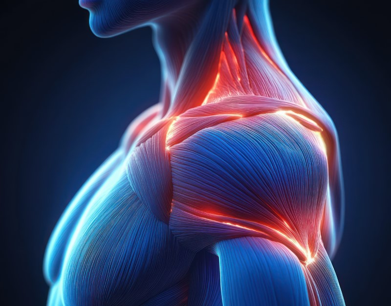
The rotator cuff is a group of 4 muscles in the shoulder, their key function is to help keep the ball of the humerus compressed in the socket. They also assist in the rotational movements of the humerus within the socket. These muscles are critical in maintaining the stability of the shoulder. They are commonly injured due to their unique anatomy and the extremely dynamic movements about the shoulder joint. They are most commonly injured where they turn into tendons and run through bony tunnels as they enter into the shoulder joint. These four muscles are called your S.I.T.S muscles; supraspinatus, infraspinatus, teres minor and subscapularis. The supraspinatus is by far the most commonly injured. These tendons can develop tendonitis or tears. These injuries can be acutely disrupted or develop chronic wear and tear.Who is at risk
If we separate these two injuries between acute tears and chronic degeneration we can better define who is at risk. In the acute tear category we have our younger adult population who can tear these tendons from overhead throwing activities or acute trauma such as a fall on an outstretched arm. We also see our elderly population suffer acute tears secondary to thinning degenerating tendons. Outside of these two populations we see an array of patients who suffer chronic wear and tear of the tendon from overhead use, poor biomechanical movements patterns about the shoulder, muscle imbalance and repetitive use.How Pt can help
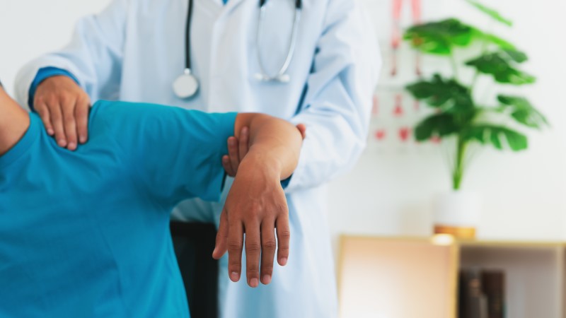
At Wall 2 Wall Sports & Rehab this is our wheelhouse, we have extensive training in the differential diagnosis of complex shoulder injuries. We have post doctoral education with world renowned shoulder and elbow mentors who have allowed us to use advanced techniques in correctly diagnosing and treating shoulder injuries. During your shoulder evaluation you can expect a comprehensive assessment and a thorough explanation of your pathology and our plan of care.Labrum
Anatomy
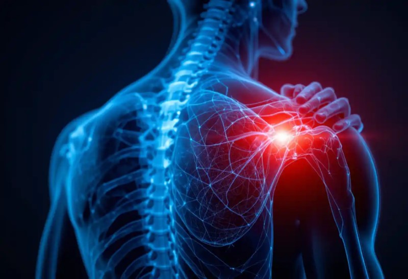
The shoulder joint is made up of two bones, the head of the humerus (ball) and the glenoid (socket), the glenoid is lined with a ligamentous type structure (labrum) that deepens the shallow socket and allows the ball to fit more concurrently in the socket. The labrum also creates negative pressure to help our shoulders stay stable. The shoulder joint is anatomically made for mobility while our hip joint is anatomically made for weight bearing. If the muscular control of the humeral head within the glenoid is not perfect, then we are at risk of fraying or tearing the labrum and or damaging our rotator cuff tendons.Who is at risk
We see a wide array of individuals with labral pain and injuries, we see a high number of overhead athletes as well as contact sport athletes with these types of injuries.How Pt can help
Physical therapy is the gold standard for labral tears in the shoulder. Unfortunately when operated on the results are varied and a lot of overhead athletes have a difficult time getting back to throwing or participating at their previous level. During your PT sessions you will focus on thoracic spine mobility, shoulder neuromuscular control and rotator cuff stability, joint mechanics and mobilization to balance the muscle tone and tightness, progressions back to throwing and sport.Instability
Anatomy
Shoulder instability can be caused by traumatic dislocation events, 95% of shoulder dislocations occur in the anterior direction, very few dislocations occur inferior or posteriorly. When the dislocation occurs the labrum can be sheared off of the bony Glenoid attachment.Who is at risk
Risk factors for shoulder dislocations are individuals who participate in extreme sports or who have recurrent repetitive microtrauma to the shoulder creating subtle underlying instability. The major cause of primary shoulder dislocation is traumatic injury. Almost 95% of first-time shoulder dislocations result from either a forceful collision, falling on an outstretched arm, or a sudden wrenching movement. In these individuals the stabilizing structures are forcefully stretched in a sudden manner. About 5% of dislocations have an atraumatic origin (eg, minor incidents such as raising the arm or moving dur- ing sleep).These individuals may have capsular laxity or al- tered muscle control of the shoulder complex or both.How PT can help
Shoulder instability is critical to be addressed early and often as if the underlying instability is left untreated you can develop early onset osteoarthritis cartilage wear and tear and lead down the pathway of a shoulder replacement. If the secondary damage from a shoulder dislocation is severe, surgical interventions are performed. There are a number of surgical procedures depending on the tissue that is damaged. ▪ Bankart Repair (Labrum) ▪ SLAP repair ▪ Biceps tenodesis/tenotomy ▪ Capsular shift ▪ Ligation/plication ▪ LatarJet ▪ Remplissage ▪ Thermal shrinkageLittle League Shoulder
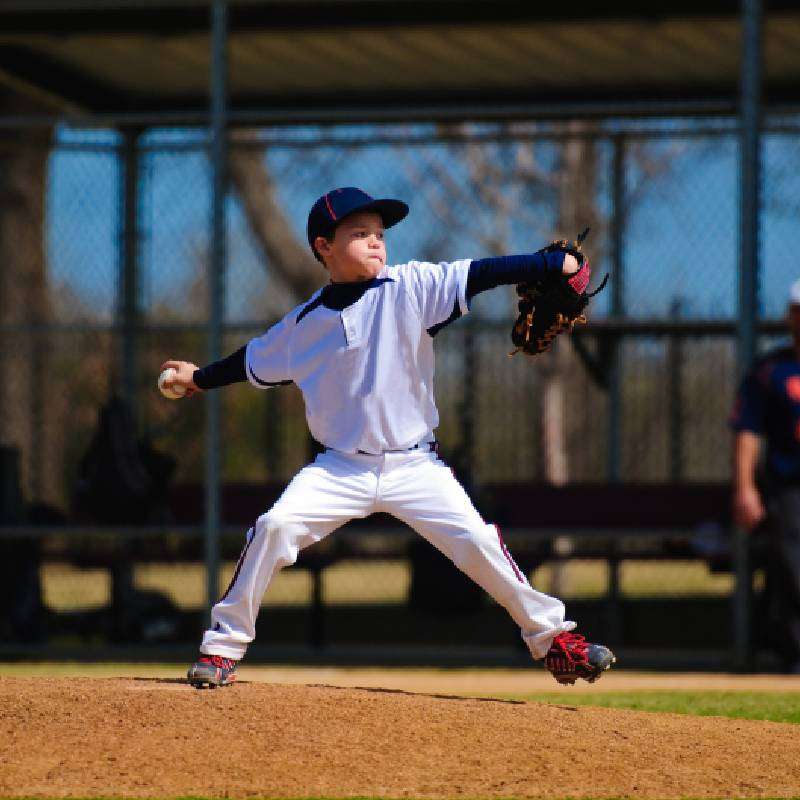
High-velocity and high-volume throwing and throwing while fatigued are significant risk factors for injury. Improved awareness and adherence to throwing guidelines should limit the number of injuries. Proper identification and treatment of injuries when they do occur can allow our skeletally immature athletes to safely return to overhead sports activities. Close to one-third of youth baseball pitchers will experience shoulder or elbow pain. This prevalence increases with age. When looking at youth baseball pitchers alone, between 26% and 35% will experience elbow or shoulder pain each year (38). Furthermore, in 2011, the results of a 10-year prospective study following 481 initially healthy youth pitchers aged 9 to 14 years demonstrated 5% incidence of serious injury resulting in surgery or retirement from base- ball (21). (Blazuk et al. 2015)